You might be wondering what X-rays are and how they came to be!
X-rays are a quick way of producing images of the inside of your body. X-rays cannot be seen or felt, but they help doctors to view the inside of your body without having to cut you open. They were accidentally discovered by a scientist called Wilhelm Röntgen in 1895. He worked at the University of Würzburg, Germany.
Why are X-rays called by this specific name?
Wilhelm Röntgen was doing tests with a cathode-ray tube when he noticed that a green glow was discharged from the tube, which was covered with a piece of black cardboard. This glow passed through the black cardboard and was projected onto a screen a few feet away. He realised that this glow was invisible rays. He named these rays “X” because they were “unknown” rays.
The early use of X-rays
Röntgen noticed that X-rays could also pass through human tissues. He then filmed the hand of his wife, Bertha, to show that X-rays could be used in medicine. The photograph of Bertha’s hand was the first photograph of a human body part taken using X-rays. Many doctors and scientists then began to investigate different parts of the body using X-rays.
an X-Ray view of the hand and wrist of a four year-old child,Credit:George Eastman
X-rays changed the world of medicine
Diverse fields benefitted from the discovery of X-rays. The field of medicine benefitted the most because radiology became a specialized field of study. X-ray imaging is also known as radiography . In today’s world, X-rays are used by doctors to treat all sorts of medical conditions in people of all ages, including children.
Who needs an X-ray?
There are different types of X-rays that doctors use to treat their patients. X-rays take images of different parts within your body, and doctors may use X-rays to check for:
• Broken bones and infections, tooth decay, and bone cancer.
• Lung infections, breast cancer, and heart failure.
• Stomach problems and evidence of swallowed items.
Are there any special things to consider before taking an X-ray?
You usually do not need to do anything special to prepare for a diagnostic X-ray . The doctor may ask you to remove jewelry or hairpins, or to stop eating or drinking several hours before doing other types of X-rays.
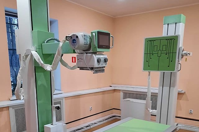
An X-ray machine is a great tool in diagnosis of diseases, Credit: Wikimedia/Oleg Bor
Are there risks associated with X-rays?
There is a far better understanding of the risks of X-ray radiation in the modern-day world than there was at first. Nowadays, scientists have developed procedures to significantly minimize the risk of overexposure to radiation during X-ray procedures. If you have any concerns about the potential risks of X-rays, you should discuss this with your doctor. You should tell your doctor if you are pregnant or suspect that you are pregnant before getting an X-ray.
X-ray technology continues to advance
Röntgen’s work earned him the first Nobel Prize in physics in 1901. This is a great achievement for an accidental discovery. The use of radiation continues to develop. X-ray technology is now widely used in devices such as airport security scanners.